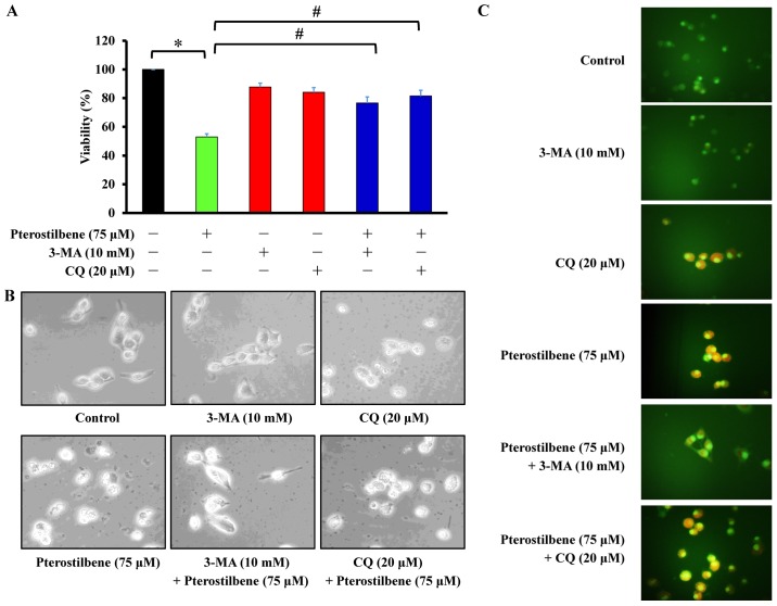
Figure 5.
Effects of the autophagy inhibitors 3-MA and CQ on the pterostilbene-induced autophagy of CAR cells. Cells were exposed to 75 μM pterostilbene for 24 h following pretreatment with 10 mM 3-MA and 20 μM CQ for 1 h. (A) Cell viability was detected using an MTT assay. Values represent the mean ± standard deviation of three independent experiments. *P<0.05 vs. untreated control; #P<0.05 vs. cells treated with pterostilbene alone. (B) The autophagic characteristic of CAR cells were observed by phase-contrast microscopy (magnification, ×400). (C) Acidic vesicular organelles indicating cell autophagy were detected using staining with acridine orange and assessed using a fluorescence microscope. Each representative image was taken from three independent experiments (magnification, ×400). 3-MA, 3-methyladenine; CQ, chloroquine.

