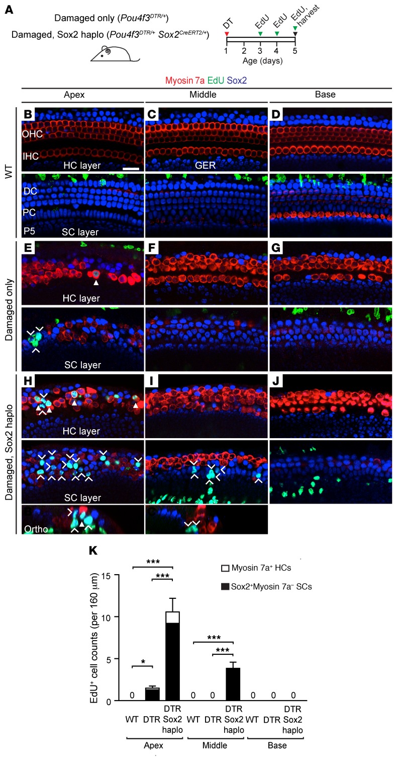
Figure 2. Reduced Sox2 levels enhance and expand the domain of proliferation in the damaged neonatal mouse cochlea.
(A) Schematic of hair cell ablation in neonatal cochlea. Briefly, Pou4f3DTR/+ and Pou4f3DTR/+ Sox2CreERT2/+ mice were injected with DT on P1 to induce hair cell loss, followed by administration of EdU (P3–P5), and cochleae were examined on P5. (B–D) No EdU+ hair cells or supporting cells were found in any of the 3 WT cochlear turns. (E–G) In the Pou4f3DTR/+ cochlea, after DT-induced hair cell damage, EdU+myosin 7a+ hair cells (arrowhead) and some EdU+Sox2+ supporting cells (chevrons) were observed in the apical turn, but not in the basal or middle turns. (H–J) In Pou4f3DTR/+ Sox2CreERT2/+ cochlea, there was robust EdU labeling of both myosin 7a+ hair cells (arrowheads) and Sox2+ supporting cells (chevrons) in the apical turn. EdU+Sox2+ supporting cells were also found in the middle turn. (K) Quantification of EdU+myosin 7a+ hair cells and myosin 7a–Sox2+ supporting cells per cochlear turn. Data represent the mean ± SD. *P < 0.05 and ***P < 0.001, by 1-way ANOVA with Holm-Sidak multiple comparisons test. n = 5–6. Scale bar: 20 μm.

