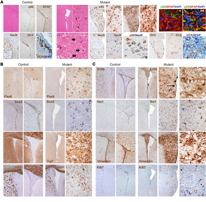
Figure 4. Targeted inactivation of Tsc1 and Pten in late-postnatal SVZ NSCs induces SEGA-like lesions.
(A) An aberrant expansion of the lateral ventricles was observed in TPN mice activated at P15–17, which were characterized by the presence of small (H&E, arrowheads) and large cells (H&E, thick arrows) intermixed within a lax fibrillary matrix (H&E; original magnification, ×40, ×400, and ×800). These structures, which were never observed in control brains (H&E; ×40), contained many pS6-IR cells (×100 and ×400). GFAP immunoreactivity was found mostly in reactive glial cells (IHC; ×200 and ×400; IF, pS6 in green, GFAP in red, and DAPI in light blue; ×400, panel with single asterisk), and less frequently in clusters of pS6-IR cells (IF, pS6 in green, GFAP in red, and DAPI in light blue; ×400, panel with double asterisk). Ectopic NeuN-IR neuronal cells were scattered throughout the lesions, some of which were pS6-IR (×200 and ×400). As opposed to SENs, only rare DCX-IR cells were found, which never colabeled with GFAP (×200 and ×400; controls, ×40, ×100, and ×400 for inset and DCX/GFAP staining). (B) Expression of Pax6 and Sox2 was restricted to large cells within the lesions, whereas Tuj1 expression was detected in most cells (mutants, ×40 and ×400; controls, ×40 and ×100). As opposed to SENs, several MBP-IR cells were found in the SEGA-like lesions. (C) S100β-IR cells were found in the intact ependymal layer (arrow), as in controls (×40 and ×100), as well as within the lesions (×40 and ×400). High frequency of Iba1-IR microglial cells and vimentin-IR reactive glial cells was observed in the lesions as compared with SENs (×40, ×200, and ×400; controls, ×40 and ×100). The mitotic index of the lesions as measured by Ki67 was still very low (×40, ×200, and ×400).

