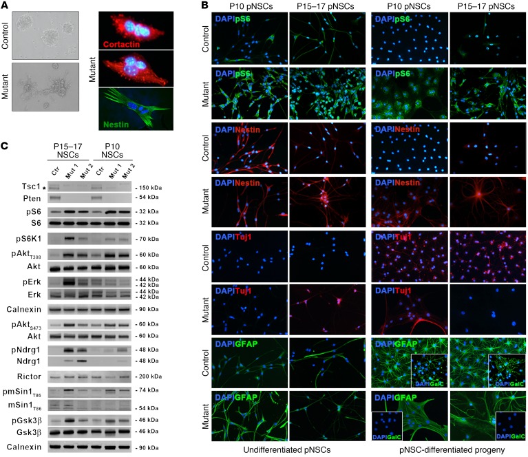
Figure 8. Postnatal NSCs isolated from SENs and SEGAs developing in TPN mice retain molecular and cellular features of their lesion of origin.
(A) Phase contrast microphotographs highlight the increased adhesive behavior of mutant pNSCs as compared with controls (representative P15–17 TPN pNSCs line L1; original magnification, ×400). SEGA-derived pNSC cultures contained multinucleated cells (representative P15–17 TPN pNSCs line L90; red, cortactin; green, nestin; ×800). (B) Both undifferentiated and differentiated P10 and P15–17 TPN cultures (L22 and L1) showed higher pS6 phosphorylation and nestin expression than controls (L26 and L4) (×400). Abnormal expression of neuronal and glial markers, such as Tuj1, GFAP, and GalC-IR, was retrieved by IF in both undifferentiated and differentiated P10 and P15–17 mutant cultures as compared with controls (×400). Data are representative of 3 pairs of pNSC lines for each activation time. (C) WB of undifferentiated P10 and P15–17 pNSCs highlighted efficient deletion of Tsc1 (*specific band) and Pten as well as different hyperactivation of mTORC1, Akt, ERK, and mTORC2 in mutant versus control pNSCs, with pERK and mTORC2 being more activated in SEGA pNSCs than in SEN pNSCs.

