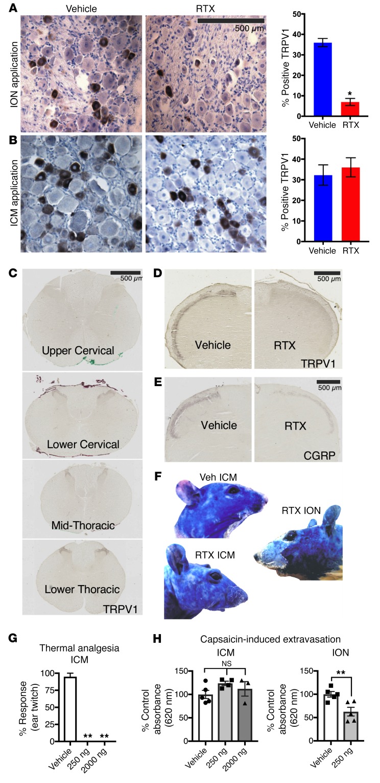
Figure 4. Effects of peripheral versus central targeting of TRPV1 on sensory ganglia.
Trigeminal ganglia sections (10 μm, paraffin-embedded) were stained for TRPV1 following 250 ng RTX or vehicle treatment. (A) There was a significant decrease (1-way ANOVA, Scheffé post hoc test, n = 6 sections, n = 3 rats, *P < 0.05) in TRPV1+ cells in the trigeminal ganglia of rats treated with RTX perineurally around the ION as compared with rats treated with the PBS-vehicle. (B) ICM administration of either RTX or PBS-vehicle did not significantly affect the proportion of cells expressing TRPV1. Counts were completed in various regions of the trigeminal ganglia (V1, V2, V3), and the proportion of TRPV1+ cells did not vary significantly. (C) ICM injection of RTX reduced TRPV1 staining in the brainstem and upper cervical spinal cord regions by at least 90% (n = 1). Injection of RTX (250 ng, 10 μl) produced loss of TRPV1+ neurons within the upper cervical region, but there was return of staining at the lower cervical/upper thoracic level and regions more caudal. (D and E) There was a strong reduction in markers of DRG neuronal axons within the brainstem following ICM treatment with RTX (n = 1). Additional quantitation is provided in Supplemental Figure 8. (F–H) Peripheral DRG function was tested by examination of capsaicin-induced plasma extravasation in trigeminally innervated regions of the face after ICM and ION RTX. While ICM RTX completely blocked thermal responses on the ear (G, **P ≤ 0.01, n ≥ 4, ANOVA, Dunnett’s post hoc), it had no effect on capsaicin-induced extravasation (H, n ≥ 3, ANOVA, Dunnett’s post hoc). In contrast, ION injection of RTX significantly blocked extravasation (P ≤ 0.05, n = 5, Student’s t test).

