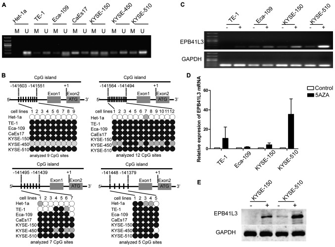
Figure 2.
Reduced expression of EPB41L3 in ESCC is due to methylation. (A) Methylation-specific polymerase chain reaction analysis of the seven cell lines revealed that EPB41L3 was partially methylated in all six ESCC cell lines while it was unmethylated in the normal Het-1a cells. (B) Representation schematic of the pyrosequencing results for the methylation status of the CpG promoter in the seven cell lines tested. Gray boxes indicate exons. Vertical bars indicate CpG sites examined for methylation. Black, gray, and white circles represent hypermethylation, partial methylation and nonmethylation, respectively. The schematic illustrates the representative results of pyrosequencing of a cytosine residue(s) at −141551, −141494, −141439 and −141379 bp of the EPB41L3 promoter. (C) Treatment with 5-Aza-2-deoxygcitidine restored the expression of EPB41L3 in ESCC lines, suggesting that downregulation of EPB41L3 in ESCC is due to methylation. (D) Reverse transcription-quantitative polymerase chain reaction analyses revealed that EPB41L3 expression was increased in ESCC cell lines following 5-Aza-2-deoxygcitidine treatment. The increase was most evident in the KYSE-510 cells. In the other cell lines, although an increase was observed, this was not as evident as in the KYSE-510 cells. (E) Western blot analyses revealed that EPB41L3 expression was increased in KYSE-150 and KYSE-510 cells following 5-Aza-2-deoxygcitidine treatment. EPB41L3, erythrocyte membrane protein band 4.1 like 3; ESCC, esophageal squamous cell carcinoma; M, methylated primer; U, unmethylated primer; 5aza, 5-Aza-2-deoxygcitidine.

