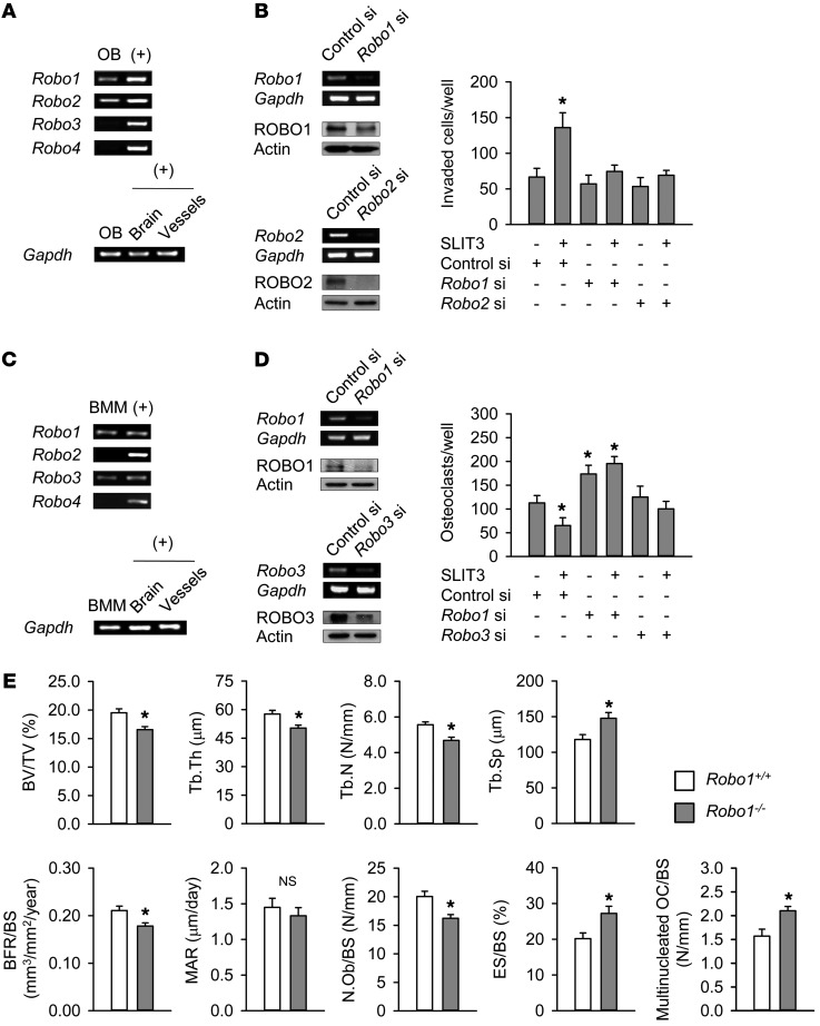
Figure 4. Osteopenic phenotypes in Robo1–/– mice.
(A) Expression of Robo mRNA in mouse calvaria osteoblasts using semiquantitative RT-PCR. Mouse brain and vascular tissues were used as positive controls (+) for Robo1–3 and Robo4, respectively. (B) Semiquantitative RT-PCR and Western blotting analysis of ROBO1 and ROBO2 after transfection with siRNA for 24 hours in mouse calvaria osteoblasts. The SLIT3-stimulated (1.0 μg/ml) directional migration of mouse calvaria osteoblasts with or without Robo1 siRNA or Robo2 siRNA was assessed using a Boyden chamber system. SLIT3 treatment was for 24 hours, after which the invaded cell numbers were counted. (C) Analysis of Robo mRNA levels in mouse BMMs using semiquantitative RT-PCR. (D) Semiquantitative RT-PCR and Western blotting of ROBO1 and ROBO3 after transfection with siRNA for 24 hours in mouse BMMs. The SLIT3-mediated (1.0 μg/ml) suppression of osteoclastogenesis with or without Robo1 siRNA or Robo3 siRNA was assessed. SLIT3 treatment was for 4 days with 15 ng/ml M-CSF and 15 ng/ml RANKL, and TRAP-positive cells with more than 3 nuclei were counted. (E) Histomorphometric analyses of the femurs of 25-week-old male Robo1–/– mice and WT littermates (n = 4–5 per group). Data are presented as mean ± SEM. In vitro experiments were performed 3–4 times independently. *P < 0.05 vs. untreated control or WT mice using the Mann-Whitney U test or Kruskal-Wallis test followed by Bonferroni’s correction.

