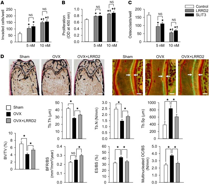
Figure 6. Effect of LRRD2 of human SLIT3 on bone mass in OVX mice.
(A) Directional migration of mouse calvaria osteoblasts upon treatment with the same molar concentration of SLIT3 (1.0 μg/ml = 10 nM) and LRRD2 for 24 hours using a Boyden chamber system. The invaded cell numbers were counted. (B) Proliferation of mouse calvaria osteoblasts in the presence of SLIT3 or LRRD2 for 48 hours assessed using a BrdU incorporation assay. (C) TRAP staining of mouse BMMs exposed to 15 ng/ml M-CSF and 15 ng/ml RANKL in the presence of SLIT3 or LRRD2 for 4 days. TRAP-positive cells with more than 3 nuclei were counted. (D) Von Kossa staining and histomorphometric analyses including calcein double-labeling of the femur of sham-operated, OVX, and LRRD2-treated OVX mice (n = 7 per group). The female C57BL/6J mice were OVX at 8 weeks of age, and 2 μg LRRD2 was injected via the tail vein twice a day (mean 0.192 mg/kg/day) from 12 weeks of age for 4 weeks. The same volume of saline was injected in the other groups. Mice were then sacrificed for analyses at 16 weeks of age. Scale bars: 500 μm (left panels); 10 μm (right panels). Data are presented as mean ± SEM. In vitro experiments were performed 3 times independently. *P < 0.05 vs. untreated control or between the indicated groups using the Mann-Whitney U test or Kruskal-Wallis test followed by Bonferroni’s correction. †P < 0.05 vs. 5 nM-treated group using Mann-Whitney U test.

