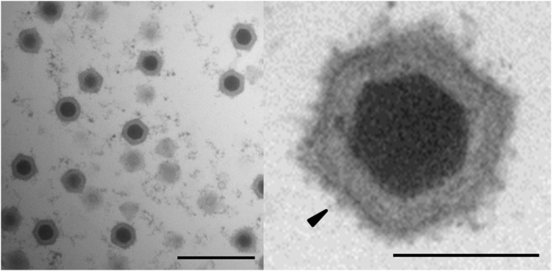
Figure 3.
Electron micrograph of virions in a diseased D. magna. Ultra-thin section of a diseased Daphnia reveals icosahedral virions consisting of an electron-dense core and an icosahedral outer structure in the host cytoplasm. High magnification of an icosahedral virion (right) reveals that the outer surface of the viral capsid is covered with fibers (black arrow). Left: bar represents 500 nm; right: bar represents 100 nm.

