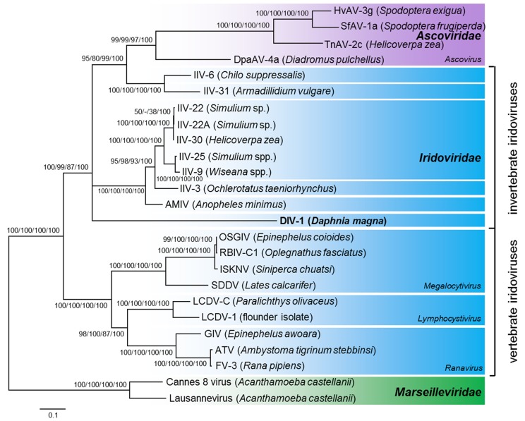
Figure 4.
Phylogenetic position of the Daphnia iridescent virus 1 using a maximum likelihood tree based on ten concatenated proteins. Maximum likelihood, neighbor joining and maximum parsimony bootstrap values (1000 replicates), and Bayesian posterior probabilities are indicated at the inner nodes. Select members of the Marseilleviridae were used as the outgroup. Hosts are given in brackets. GenBank/EMBL/DDBJ accession numbers are given in Table S1.

