
Figure 4.
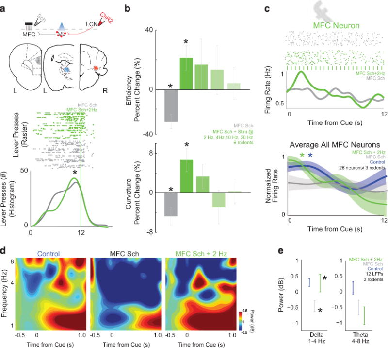
Stimulation of cerebellar projections can compensate for frontal dysfunction. We modeled aspects of frontal dysfunction in schizophrenia by pharmacologically disrupting medial frontal cortex (MFC) D1 dopamine receptors with SCH23390. (a) Histological analyses revealed electrode infusion cannula ensemble placement in the MFC (gray), optical cannula in the ventrolateral thalamus (VL, blue) and channelrhodopsin2 (ChR2) infusion/cannula placement in the lateral cerebellar nuclei (LCN) for individual animals as represented by each dot. Stimulating LCN-VL projections at 2 Hz could rescue behavioral deficits caused by MFC SCH23390 (lower plot in a; MFC SCH23390: gray— LCN-VL stimulation: green; ticks are individual lever presses from the entire experiment). (b) Group data from eight rodents showed impaired behavior with MFC SCH23390 and behavioral rescue with 2 Hz stimulation of LCN-VL projections. (c) During interval timing, MFC neurons have time-related ramping, which is decreased with MFC SCH23390 (gray). For this neuron, with LCN-VL 2 Hz stimulation, ramping increased (green); each blue tick is a 473 nm laser pulse. With LCN-VL 2 Hz stimulation, ramping activity increased as measured by average activity or by linear regression; MFC-saline without stimulation is labeled in blue. Data from 26 MFC neurons in three rodents; asterisk indicates significance at P<0.05 via χ2 test. (d) Similar to humans, rodents have a burst of delta/theta activity as shown by time-frequency analyses, which is decreased in animals with MFC D1DR blockade (gray—MFC SCH23390; n = 12 LFPs in three rodents). (e) With LCN-VL 2 Hz stimulation (green), delta MFC activity significantly improved. Asterisks indicate significance at P<0.05 via paired t-test displayed; all data are displayed as mean ± s.e.m.