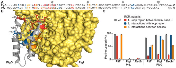
Figure 4.
PCP loop 1 modification alters interactions with homologous A domains. Residues were mutated in loop 1 (red), underneath loop 1 (blue), and between helix I and II (orange). (A) Sequence alignment of PigG and PltL using MUSCLE.35 (B) holo-PigG NMR structure docked to model structure of PigI. Mutated residues are highlighted in red, blue, and orange and are explained in (C). (C) Aminoacylation assays with mutated PigG and either PigI or PltF. Astericks indicate PltL mutant 2 species was not analyzed due to instability.

