FIGURE 10.
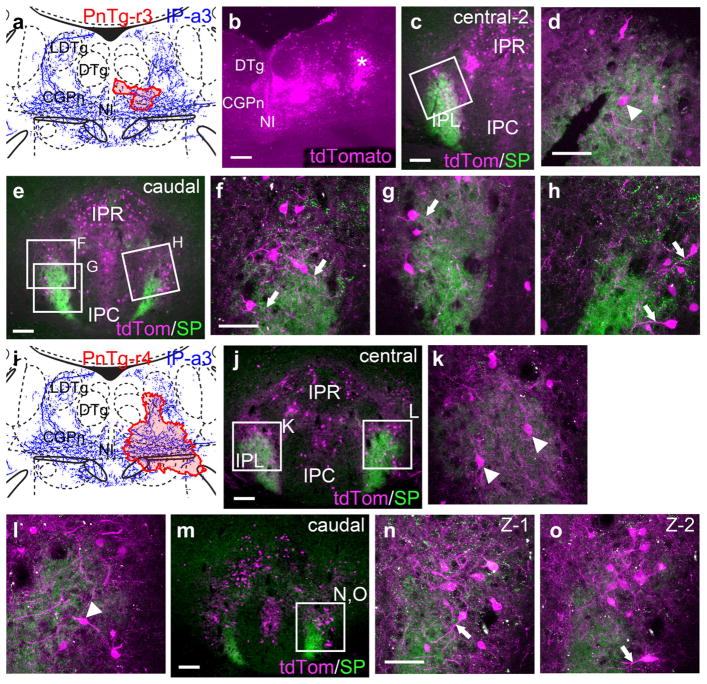
Interpeduncular nucleus projections to the lateral pontine tegmentum. In order to effectively label IP neurons and their processes, a transgenic reporter line ROSA-lsl-tdTomato (Ai14) was used. tdTomato expression was induced by retrograde transport of Cav2-Cre virus injected into the pontine tegmentum. (a) Map of the Cav2 injection site (red shaded area) for case PnTg-r3, labeling a small area of the CGPn. The diagram corresponds to bregma −5.52 in a standard atlas. (b) Cre-induced expression of tdTomato at the site of the Cav2-Cre injection. Labeled cells far lateral to the injection site (asterisk) probably represent retrograde labeling within the plane of the section, rather than spread of the injected virus. (c, d) Low power (c) and confocal (d) images of the central IP in case PnTg-r3. A labeled cell body within the IPL is indicated by the arrowhead. (e–h) Low power (e) and confocal (f–h) images of the caudal IP in case PnTg-r3. Neural processes (probably dendrites) that extend into the habenulo-recipient area of the IPL from its periphery are marked with arrows. (i) Map of the Cav2-Cre injection site (red shaded area) for case PnTg-r4, labeling a more extensive area of the CGPn and adjacent structures. (j–l) Low power (j) and confocal (k, l) images of the central IP in case PnTg-r4. (m–o) Low power (m) and confocal (n, o) images of the caudal IP in case PnTg-r4. (n, o) show two confocal Z-stacks acquired at different depths of the same region boxed in (m). Scale: (b, c, e, j, m), 100 μm; (d, f, k, n), 50 μm [Color figure can be viewed at wileyonlinelibrary.com]

