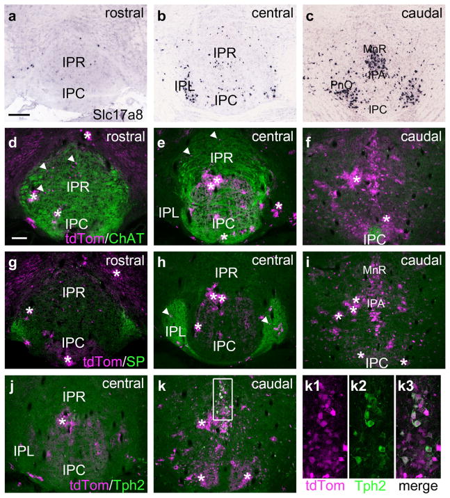
FIGURE 13.
Slc17a8 (Vglut3) expression in the interpeduncular nucleus. (a–c) In situ hybridization for Slc17a8 mRNA expression in the IP (Allen Brain Atlas case # 71587918). Results are generally concordant with the pattern of expression using a Slc17a8Cre driver in the subsequent panels. In (b) more Slc17a8 cells are observed in the IPL than expected based on the Cre-driver results. Since no specific marker is available for the IPL in the in situ data, the exact plane of section cannot be determined, and these Vglut3 neurons may lie caudal to the IPL. (d–f) Expression of a tdTomato reporter in a Slc17a8Cre/Ai14 mouse, immunostained for ChAT to reveal vMHb afferents. Slc17a8Cre-driven reporter expression was generally consistent with Vgllut3 mRNA expression, however ectopic expression was observed in sporadically distributed astrocytes (asterisks), which were easily recognized by their patchy morphology. Only rare Slc17a8-expressing neurons were observed in the area of IPR and IPC receiving vMHb fibers, examples are shown by arrowheads in (d, e). In the caudal IP Slc17a8 neurons were abundant, but only caudal to the habenulorecipient areas. (g–i) Sections adjacent to those in (d–f) immunostained for SP to show dMHb afferents. (j–k) Sections adjacent to those in (g–i) immunostained for Tph2 to reveal 5HT-expressing neurons. Confocal images in (k1–k3) show co-localization of tdTomato and Tph2 in slc17a8Cre neurons. Scale: (a) 200 μm; (b) 100 μm [Color figure can be viewed at wileyonlinelibrary.com]

