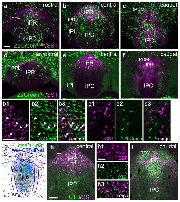
FIGURE 14.
Somatostatin expression defines a subset of GABAergic projection neurons in caudal IPR. (a–c) A Gad2cre driver line was interbred with the reporter line Ai6 in order to label all GABAergic neurons with the marker ZsGreen. ZsGreen was colocalized with SST by immunofluorescence in the rostral (a), central (b) and caudal (c) IP. SST+ IP neurons define a caudal division of IPR. A small number of SST+ cell bodies are also found in IPC and IPL. 35/37 of the SST+ neurons in IPR in the section shown in (b) co-expressed ZsGreen. Images (b1–b3) show confocal detail of the area boxed in (b). Arrowheads indicate examples of SST+ GABAergic cell bodies with clear nuclear outlines. (d–f) A Slc17a6Cre driver line was interbred with the reporter line Ai6 in order to label all glutamatergic neurons with the marker ZsGreen. ZsGreen was colocalized with SST by immunofluorescence in the rostral (d), central (e) and caudal (f) IP. 0/61 SST+ neurons in IPR in the section shown in (e) expressed ZsGreen. Images (e1–e3) show confocal detail of the area boxed in (e). (g–i) Retrograde tracing of efferent projections of SST+ IPR neurons. (g) Map of the CTB injection site for case MnR-r1, a midline injection encompassing much of MnR. The diagram corresponds to bregma −4.60 in a standard atlas. (h) CTB labeling in the central IP in case MnR-r1. Confocal images (h1–h3) of the boxed area show that numerous SST+ neurons in IPR are retrogradely labeled at this level. (i) CTB labeling in the caudal IP. IPDL exhibits strong retrograde labeling but does not express SST. Scale: (a, h) 100 μm; (b1, h1), 50 μm [Color figure can be viewed at wileyonlinelibrary.com]

