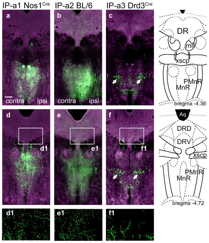
FIGURE 2.
Anterograde labeling of IP projections to the mesencephalic raphe. IP efferents to the midbrain were examined in each of the cases described in Figure 1. (a–c) IP efferents to the MnR/PMnR. The standard atlas coordinate is bregma −4.36. (d–f) IP efferents to the MnR/PMnR and DR. The standard atlas coordinate is bregma −4.72. Some loss of resolution (blurring) in the ventral part of (b) and (e), and also in Figure 3b, represents a technical issue with automated image acquisition and not an actual difference in the distribution of the signal. Arrows in (c, f) indicate bundles of fibers that have the typical appearance of fibers of passage, without varicosities. In (d1–f1) segmented views are shown, in which the local fluorescence signal has been extracted from the digital images to show detail. Scale: (a), 200 μm [Color figure can be viewed at wileyonlinelibrary.com]

