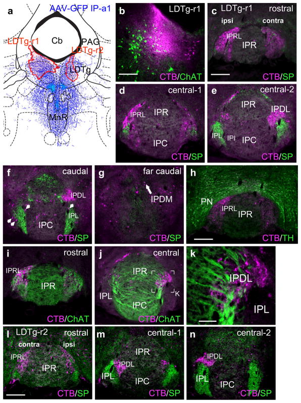
FIGURE 8.
Interpeduncular nucleus projections to the lateral dorsal tegmental nucleus. (a) Map of the CTB injection sites (red shaded areas) for two cases labeling the LDTg, in opposite hemispheres. The diagram corresponds to bregma −5.02 in a standard atlas. The pattern of anterograde tracing in IP-a1 is shown in blue. (b) Detail of injected area in case LDTg-r1, showing overlap of injected area with ChAT-expressing cholinergic neurons in LDTg. (c–g) Retrograde labeling of the IP subnuclei in case LDTg-r1 at the designated levels. Labeling predominates in IPRL, ipsilateral to the injection, and IPDL contralateral to the injection. A small number of labeled neurons are located on the periphery of SP-labeled afferent fibers in IPL bilaterally (arrows, f). (h) Retrograde labeling of the rostral IP in case LDTg-r1. TH staining reveals dopaminergic neurons of the PN, which does not overlap the retrograde labeling within IPRL. (i–k) Relationship of retrograde labeling in case LDTg-r1 to ChAT-labeled afferent fibers in IPR, IPC, and IPDL. A confocal detail image of the boxed area in (j) appears in (k). (l–n) Retrograde labeling of the IP subnuclei in case LDTg-r2. In this case, injected closer to the midline, IPRL is labeled bilaterally (l), while IPDL labeling is still predominantly contralateral (m, n). Scale: (b, c, h, l), 200 μm; (k), 50 μm [Color figure can be viewed at wileyonlinelibrary.com]

