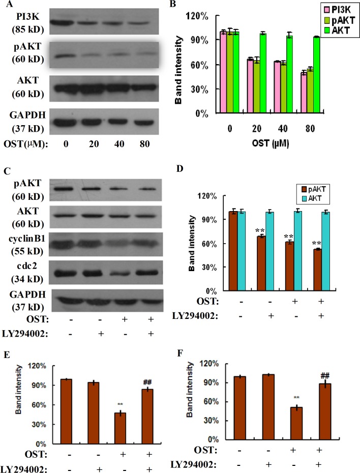
Fig 4. Osthole represses PI3K-AKT signaling in gastric cancer cells.
HGC-27 cells were treated with or without osthole for 48 h and the cell lysates were subjected to sodium dodecyl sulfate-polyacrylamide gel electrophoresis and analyzed by western blot assay (A). The expression of PI3K, pAKT and AKT proteins was determined by western blot assay. Densitometric analyses of PI3K, pAKT and AKT proteins were expressed as the mean ± SD from three independent experiments (B). HGC-27 cells were pretreated with PI3K specific inhibitor LY294002 for 2 h and followed by the indicated concentration of osthole, the cell lysates were subjected to sodium dodecyl sulfate-polyacrylamide gel electrophoresis and analyzed by western blot assay. The expression of pAKT, AKT, cyclin B1, and cdc2 proteins was determined by western blot assay (C). Densitometric analyses of these proteins were expressed as the mean ± SD from three independent experiments (D: pAKT, AKT; E: cyclin B1; F: cdc2). **P<0.01 vs. control; ## P<0.01 vs osthole group.

