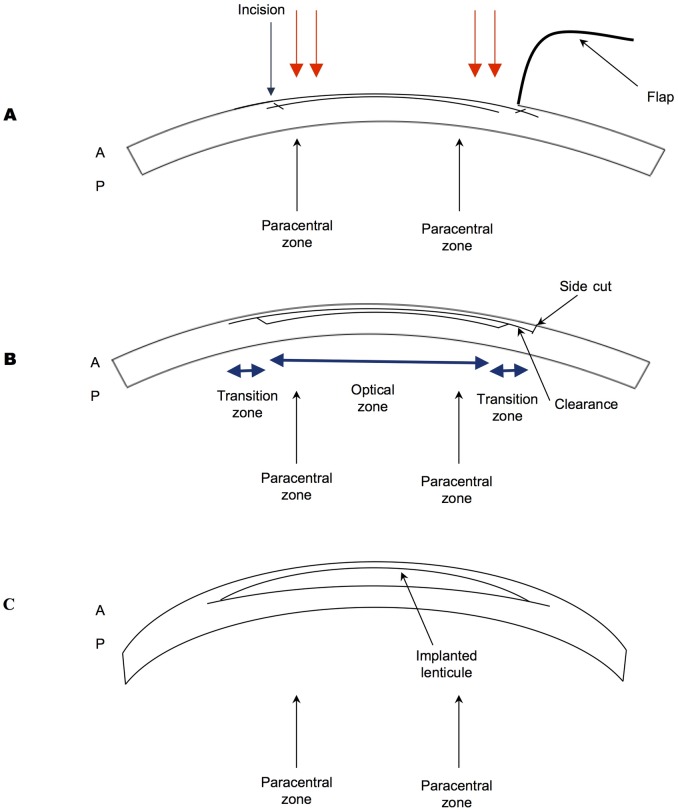
Fig 1. Cartoon schematic of hyperopic treatments.
A, LASIK: Corneal is lifted, excimer laser (represented by red arrows) is applied to the peripheral cornea in a circular pattern. B, SMILE lateral view of cornea showing optical (anterior cap) zone (5.5mm diameter), the transition zone (1mm wide) and the 90 degrees lenticule side cut C, Lenticule shape: Showing the implanted lenticule. A = Anterior Cornea, P = Posterior Cornea.

