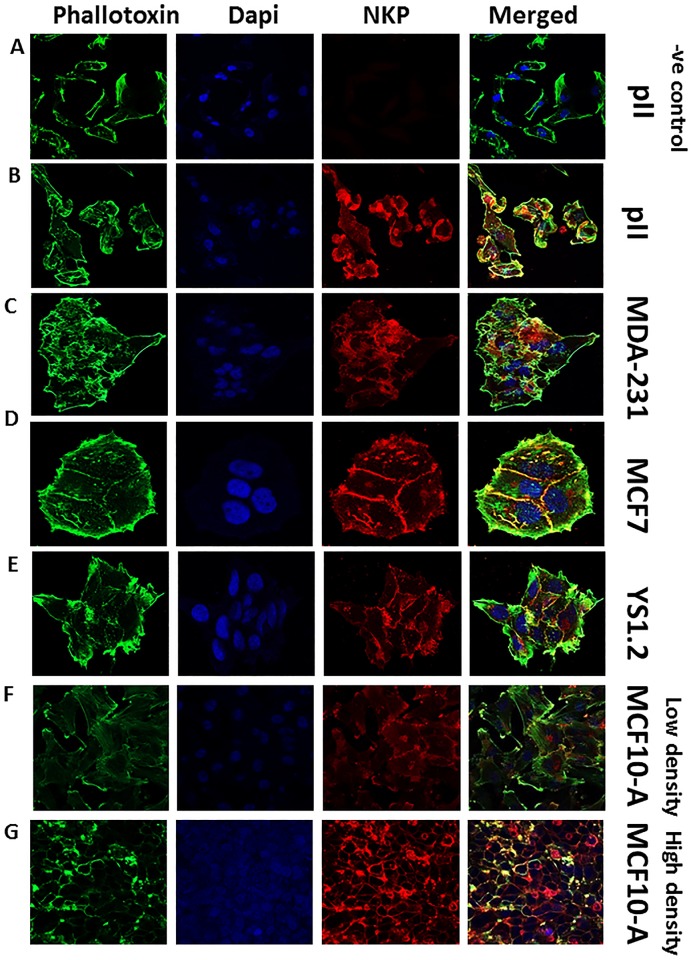
Fig 3. NKP localization in normal and breast cancer cell lines.
Cells were seeded into 8-chambered slides and allowed to grow for 48 h at 37o C/ 5% CO2. Cells were then fixed and stained with NKP antibody (red), phallotoxin (green) and DAPI (blue). Panel A represents a negative control where pII cells were incubated with secondary antibody only.

