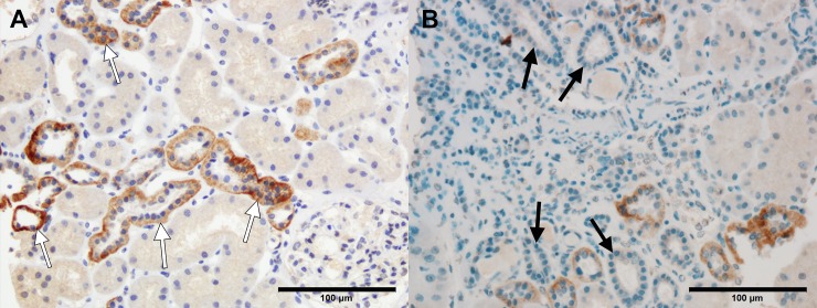
Fig 7. Representative images of immunohistochemistry (IHC) staining for klotho based on adjacent pathological features.
(A) Distal tubules surrounded by normal tubuloinsterstitium showed strong IHC staining for klotho (white arrows). (B) Distal tubules surrounded by inflamed interstitium and atrophic tubules showed absent or weak IHC staining for klotho (black arrows). Scale bar = 100 μm.

