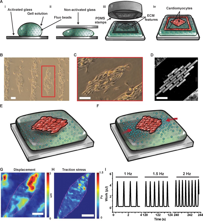
Fig 1. Micropatterned cardiac tissues on soft gels for traction force microscopy.
(A) We cast PA gels (i) sandwiching the pre-polymer solution between activated and non-activated glass (ii) before stamping fibronectin (iii) to promote cell adhesion on the gel surface (iv). With this versatile method, we engineered neonate rat ventricular myocytes (NRVM) into diamond-shaped mini tissues (B) featuring cells aligned along the major axis of the diamond (C) thanks to a micro-contact printed brick-wall pattern of fibronectin (D). By tracking the displacement of fluorescent beads embedded in the in the soft gel during cell relaxation (E) and contraction (F), we used traction force microscopy to obtain displacement (G), and stress (H) maps at the tissue level. Importantly, since we could electrically stimulate the diamond-shaped tissues, we could measure displacement, stress, and contractile work as a function of beating frequency (I). Scale bars: 100 μm.

