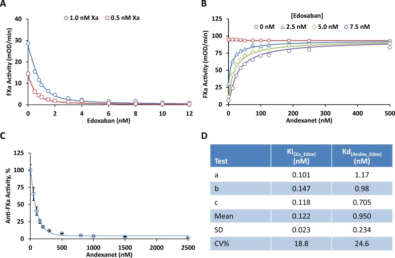
Fig 2. Edoxaban anti-FXa activity and andexanet effects in vitro.
(A) Inhibition of FXa chromogenic activity by edoxaban in a buffer system with purified enzyme. Human FXa at 0.5 nM (□) and 1.0 nM (○) were pre-incubated with increasing concentrations of edoxaban (0, 0.5, 0.8, 1, 1.5, 2, 3, 4, 6, 8, 10, 12 nM) at RT for 2 hrs. Residual FXa activity was determined by measuring the initial rates of peptidyl substrate hydrolysis (mOD405/min) at RT over 5 min. The initial velocity was fitted with Dynafit to obtain the kinetic parameters Ki and kcat using the pre-determined Km (82.2 μM) as described in Materials and Methods. Fig 2A shows a representative result from one of the three experiments shown in Fig 2D (Test a for Ki). The symbols represent the measured mean initial rate from quadruplicate wells at each edoxaban concentration. The solid lines were drawn using the best fitted values with Ki = 0.101 nM, kcat = 156 1/s. (B) Reversal of edoxaban-induced inhibition of FXa chromogenic activity by andexanet in a buffer system with purified enzyme. Human FXa (3.0 nM); different concentrations of edoxaban at 0 (□), 2.5 (△), 5.0 (◇),and 7.5 nM (○); and increasing concentrations of andexanet (0, 12.5, 25, 37.5, 50, 75, 100, 125, 188, 250, 500 nM) were pre-incubated at RT for 2 hrs. Residual FXa activity was determined by measuring the initial rates of peptidyl substrate hydrolysis (mOD405/min) at RT over 5 min. The initial velocity was fitted with Dynafit to obtain the kinetic parameters Kd and kcat using the pre-determined Km (82.2 μM) and Ki (0.122 nM) as described in Materials and Methods. Fig 2B shows a representative result from one of the three experiments shown in Fig 2D (Test b for Kd). The symbols represent the measured mean initial rate from duplicate wells at each andexanet concentration. The solid lines were drawn using the best fitted values with Kd = 0.98 nM, kcat = 176 1/s. (C) Reversal of edoxaban-induced anti-FXa activity by andexanet in human plasma. Edoxaban (76 ng/mL, 0.136 μM) and increasing concentrations of andexanet (0, 0.05, 0.1, 0.15, 0.2, 0.3, 0.5, 0.8, 1, 1.5, 2.5 μM) were prepared in human plasma and pre-incubated at RT for 30 min. Residual anti-FXa activity for edoxaban was measured as described in Materials and Methods. Fig 2C shows edoxaban anti-FXa activity (%) after normalization of the results to the mean anti-FXa value at 0 nM andexanet. Data were plotted as the mean ± standard deviation from three separate experiments. (D) Constants for edoxaban interaction with FXa (Ki) and andexanet (Kd) determined by the kinetic measurements as described in panel (A) and panel (B), respectively.

