
Figure 1. The organized alignment of polarized spermatids across the plane of the seminiferous epithelium in adult rat testes, supporting the concept of spermatid planar cell polarity (PCP) during spermiogenesis.
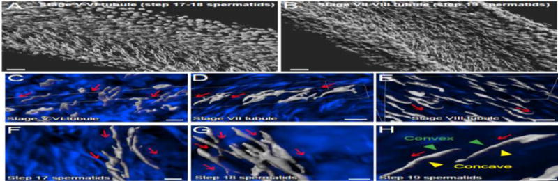
Isolated rat seminiferous tubules were obtained from adult testes as earlier described [84], stained with DAPI (4′,6-diamdino-2-phenylindole) and images were obtained by confocal microscopy, illustrating spermatid PCP across the seminiferous epithelium (A–B). Images were obtained using an inverted Zeiss LSM 880 NLO laser scanning confocal/multiphoton microscope (Carl Zeiss MicroImaging, Thornwood, NY). Optical sections of 20–100 μm of the seminiferous tubule were collected at 0.83-μm intervals along the z-axis to obtain Z-stack image series. Images were then deconvoluted using Autoquant X deconvolution software (Media cybernetics), and 3D images were subsequently obtained using Imaris (Bitplane) software package (Version 8.9), but using only the DAPI channel to show the alignment of polarized spermatid heads across the plane of seminiferous epithelium. White pseudo color was applied to the DAPI channel to obtain optimal image contrast (A–B). For other tubule images shown in C–H, deconvolved images of the Z-stack series were reconstructed in Imaris with the blue channel for DAPI. Red arrows annotate the directional (head-tail polarity) orientation of spermatid heads by pointing toward the basement membrane, also noted are the multicellular polarity in C–H. As noted in H, the convex (dorsal) and concave (ventral) sides of the spermatid head were also annotated. Scale bar, 100 μm in A, B; 15 μm in C–E; 3 μm in F–H.