
Figure 4. Malacidin mode of action.
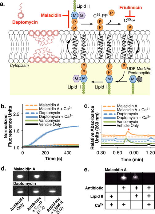
(a) Cartoon showing modes of action of daptomycin, friulimicin and malacidin. (b) In contrast to daptomycin, malacidin A does not cause MRSA membrane leakage in a SYTOX Green fluorescent assay. Error bars represent the standard deviation across three biological replicates (n = 3). (c) As seen with the cell wall biosynthesis inhibitor vancomycin, exposing MRSA to malacidin A results in the accumulation of the cell wall intermediate UDP-MurNAc-pentapeptide. The UDP-MurNAc-pentapeptide peak ([M-H]- = 1148.35) is indicated with a red asterisk on the UPLC- trace. Chromatograms are representative of at least three independent experiments. (d) Interaction of malacidin A and daptomycin with purified cell wall precursors. An interaction is indicated by a reduction of the amount of free antibiotic (visible on the thin-layer chromatography by UV light). (e) The interaction of malacidin A to cell wall precursor, lipid II, is calcium-dependent. Both TLCs in (d) and (e) are representative of at leas two replicate experiments.