Fig. 5.

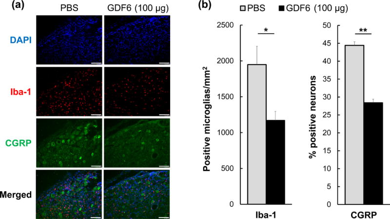
Dorsal root ganglion (DRG) immunohistochemistry in the nude rat xenograft radiculopathy model. The effect of degenerated nucleus pulposus tissues from either phosphate-buffered saline (PBS) control or 100 μg/disc of growth differentiation factor-6 (GDF6) injected rabbit intervertebral discs on the immunohistochemistry of rat DRGs in the rat xenograft model. a. Representative images of immunofluorescence for 4′,6-diamidin-2-phenylindol (DAPI) (blue), ionized calcium binding adaptor molecule-1 (Iba-1) (red), calcitonin gene-related peptide (CGRP) (green), and merged signals in experimental and control DRGs. Bar scale is 100 μm. b. On day 21 after xenograft, the number of Iba-1-positive microglia/mm2 and percentage of CGRP-positive neurons in DRGs of PBS and GDF6-injected discs transplanted onto nude rat DRGs are shown. Data are expressed as the mean ± standard error (n = 5). Unpaired t-test was used. *p < 0.05, **p < 0.01.
