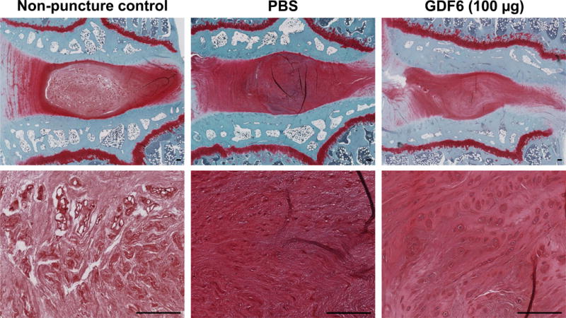
Fig. 9.

Cell morphology changes 16 weeks after anular-puncture (12 weeks after injections of phosphate-buffered saline [PBS] or growth differentiation factor-6 [GDF6]) in lumbar discs in the rabbit anular-puncture model. On safranin-O stained sections of control (non-puncture), PBS-, and GDF6 100 μg-treated discs, cell clones were not found in non-puncture and PBS-treated discs, while chondrocyte-like cell clone formation was found in GDF6 100 μg-treated discs (37.5%). Bar scale is 100 μm.
