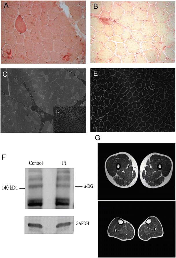
Fig. 1.

(A, B) Histology of skeletal muscle from the patient. Hematoxylin eosin staining showed moderate fiber size variability with several hypotrophic fibers and some hypercontracted fibers (A). The staining for acid phosphatase was increased (B). (C–E) Immunofluorescence with IIH6C4 antibody showing profound reduction of immunodetectable glycosylated α–DG (C) in comparison to control muscle stained with the same antibody (D). In (E) immunofluorescence for caveolin-3, showing normal protein expression. (F) Western blotting showed an expected band of 140 kDa with a 15% reduction in glycosylated α-DG compared to control. Anti-α-Dystroglycan, clone IIH6, monoclonal antibody (Millipore, Temecula, CA) was used at 1:100 dilution, according to manufacturer’s instruction. GADPH expression is used as control for protein loading. Band intensities were evaluated by densitometry using the ImageQuant 350 system (Amersham Biosciences). (G) Muscle MRI performed at 21 years disclosed a moderate fatty infiltration in muscles of the posterior thigh (adductor magnus hamstrings, and partially sartorius) and minimal fatty infiltration of soleus muscle at leg level
