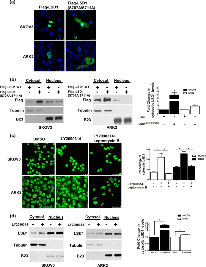
Fig. 4. Phosphorylation of LSD1 alters its subcellular localization.
a The subcellular localization of Flag-wild-type LSD1 or Flag-double-mutant (S707A/S711A) LSD1 (green) was investigated with confocal microscopy. b Nuclear and cytoplasmic fractions obtained from cancer cells (SKOV3 and ARK2) overexpressing Flag-wild-type LSD1 or Flag-double-mutant (S707A/S711A) LSD1 were analyzed with Western blot using anti-Flag, anti-tubulin (cytoplasmic marker), and anti-B23 (nuclear marker) antibodies, respectively. c Confocal microscopy image indicated the intracellular localization of endogenous LSD1 (green) in cells treated with DMSO or LY2090314 (10 µM) for 24 h or LY2090314 (10 µM) with leptomycin B (10 nM) for 24 h. Cytosolic LSD1 were quantified with MetaMorph software. d SKOV3 and ARK2 cells were treated DMSO or LY2090314 (10 µM) for 24 h. Protein levels of the nuclear or cytoplasmic fractions of LSD1, tubulin, and B23 were determined with Western blot. Results shown were obtained from three independent experiments and presented as mean ± standard error (SE). *P < 0.05, Student’s t-test

