Fig. 2.
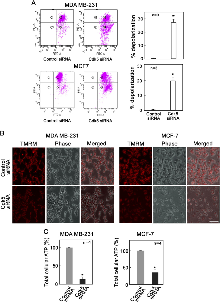
Loss of Cdk5 induces mitochondrial depolarization in breast cancer cells. Analysis of mitochondrial membrane potential using the fluorescent probes, JC-1 and tetramethylrhodamine methyl ester perchlorate (TMRM), in MDA MB-231 and MCF-7 cells depleted of Cdk5. After 72 h of transfection with Cdk5 siRNA, cells were stained with (a) JC-1 dye (5 µM) or (b) TMRM (5 µM) for 30 min at 37 °C and analyzed by flow cytometry and fluorescence live cell microscopy, respectively. Graphs in (a) represent the percent increase in number of cells with depolarized mitochondria based on the fluorescence intensity of JC-1 in Q4 of the flow cytometry data on the left panel. b Fluorescence microscopy of MDA MB-231 and MCF-7 cells probed with TMRM using an Olympus 1 × 71 microscope at ×160 magnification. Scale bar = 100 µm. Data represent one of three experiments showing similar staining pattern. c Total cellular ATP level in MDA MB-231 and MCF-7 cells was measured by bioluminescence assay. Briefly, following incubation of cell lysates with reaction buffer containing DTT, luciferin, and luciferase, luminescence was measured at 560 nm using a plate reader. Values from three (a) or four (c) independent experiments are expressed as means ± SEM. * indicates p < 0.05 using the Student’s t-test (unpaired)

