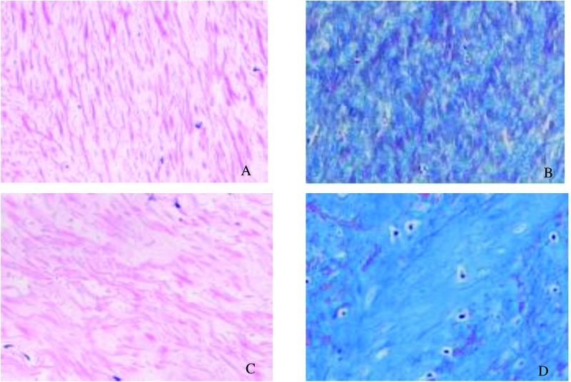
Figure 1. Histological analysis of LF specimens.
(A) In LF from the LSCS group, a large area was stained pink with a regular arrangement (H&E staining, ×200). However, in the LDH group (C), elastic fibers were disorganized and focally lost. Grading of LF fibrosis by Masson’s trichrome staining (B and D). The blue color indicates collagen fibers and the pink color indicates elastic fibers. (B) Grade 1 showed a large area was stained pink (×200). (D) In Grade 4, blue stained most of the area, indicating fibrotic change (×200).

