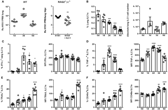
Figure 1.
H1N1 infection leads to enhanced ILC1 activation and functionality. Wild-type mice and RAG2−/−γc−/− mice were infected i.n. with 2 × 10³ ffu of the H1N1 PR8 strain. (A) Scatter plots represent viral loads of wild-type mice (left panel) and RAG2−/−γc−/− mice adoptively transferred with 2 × 10⁵ in vitro-generated ILC1s/animal (right panel; n = 4–8) as determined by foci assay and depicted as ffu per lung at the indicated time points. Lymphocytes derived ex vivo from lungs of infected wild-type mice were incubated at 37°C in medium containing brefeldin and monensin for 3 h prior to flow cytometry staining of markers related to ILC1 activation and functionality. (B) Frequencies and absolute numbers of lung-derived ILC1s. Frequencies and mean fluorescence intensities (MFI) of (C) IFN-γ, (D) TNF-α, (E) TRAIL, and (F) CD49a expressing lung ILC1s. Shown is one out of three independent experiments (n = 4–8). Bars with scatter plots represent MFI and range in frequencies with the horizontal line drawn at the mean. Asterisks denote significant values as calculated by nonparametric Mann–Whitney’s test (viral loads) or One-way ANOVA (frequency and MFI) as compared to uninfected samples; ****p ≤ 0.0001; ***p ≤ 0.001; **p ≤ 0.01; *p ≤ 0.05.

