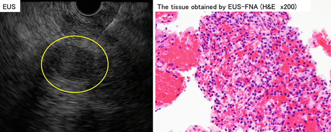
Figure 5.
Left: A low echoic lesion of 2 cm in length in the pancreatic tail was detected by EUS (yellow circle). The lesion did not include a cystic lesion. It was punctured with a 22-gauge needle and tumor tissue was collected. Right: Hematoxylin and Eosin staining showed lymphocytes, as is observed in splenic tissue. EUS: endoscopic ultrasound, EUS-FNA: endoscopic ultrasound-guided fine-needle aspiration

