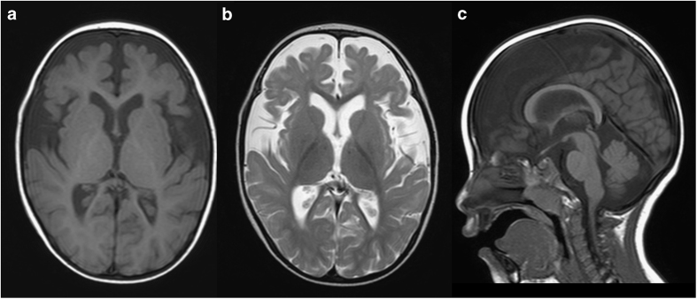
Figure 1.
Brain magnetic resonance imaging examination at 2 years of age. Axial T1- and T2-weighted images (a and b, respectively) and a sagittal T1-weighted image (c). Decreased volume of the cerebrum (a, b) and delayed myelination in the deep white matter (b) are evident. The volume of the corpus callosum is also reduced (c).

