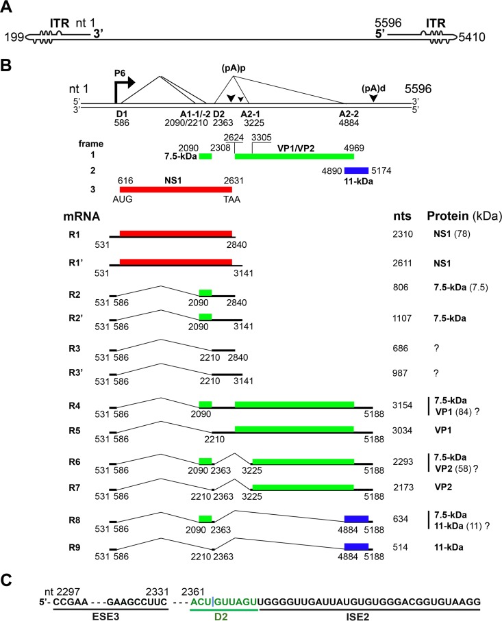
FIG 1.
B19V transcription map. (A) B19V genome. The linear single-stranded B19V genome is shown in the negative sense, with unpaired or mismatched bases diagrammed as bulges and bubbles. ITR, inverted terminal repeat. (B) Transcription profile. The B19V duplex genome is shown at the top. P6 represents the viral promoter, D1 and D2 denote splice donor sites, and A1-1, A1-2, A2-1, and A2-2 denote splice acceptor sites. Different open reading frames are shown by different colors (red, green, and blue). (pA)p and (pA)d represent polyadenylation sites at the proximal and distal ends, respectively. At the bottom, 9 major RNAs encoding different viral proteins, as indicated, are shown. Question marks indicate that it is unknown whether or not the protein is expressed from the species of mRNA. (C) ESE3, D2, and ISE2 elements. The donor 2 (D2) site of the B19V pre-mRNA is flanked by exon splicing enhancer 3 (ESE3) on the left and intronic splicing enhancer 2 (ISE2) on the right. They act as cis-acting elements for splicing at the D2 site.

