FIG 9.
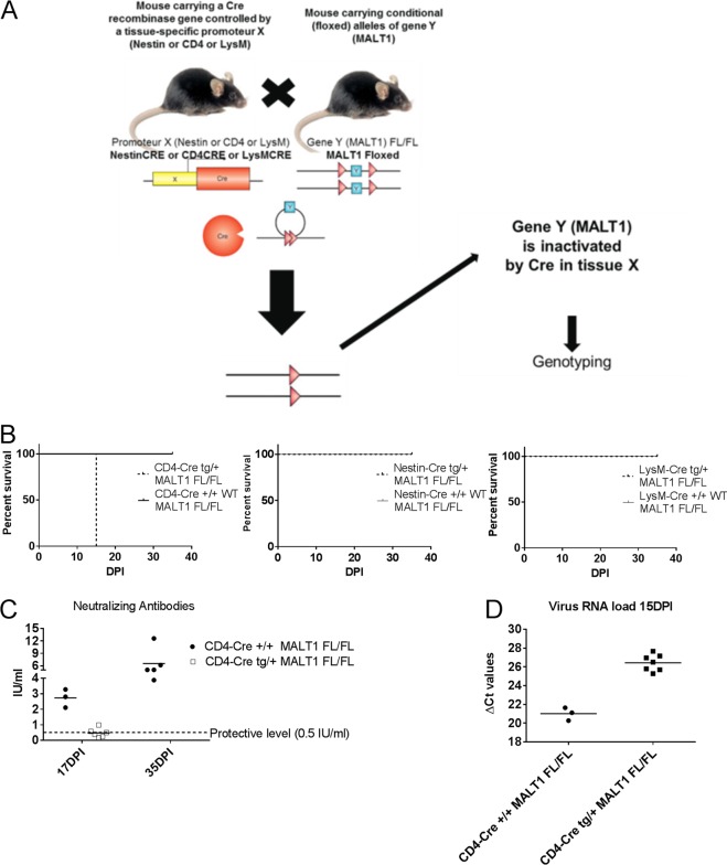
Impact of specific MALT1 deficiency in T cells, myeloid cells, or cells from neuroectodermal origin on ERA virus infection. (A) Schematic overview of conditional MALT1−/− mouse generation. Mice expressing the CRE recombinase gene under the influence of the CD4, LysM, or Nestin promoter were crossed with MALT1FL/FL mice to generate conditional mice lacking MALT1 in T cells, myeloid cells, or cells from a neuroectodermal origin, respectively. Conditional mice and their wild-type littermates were genotyped and selected for each experiment. (B) Survival of infected mice. Mice were infected intranasally with ERA virus and monitored for disease development and survival. Mice lacking MALT1 in cells from neuroectodermal origin (Nestin-Cretg/+ MALT1FL/FL) and myeloid cells (LysM-Cretg/+ MALT1FL/FL) developed only mild disease, comparable to their wild-type littermates. Mice lacking MALT1 in T cells (CD4-Cretg/+ MALT1FL/FL) presented the same phenotype as the full MALT1−/− and developed severe disease requiring euthanasia at 15 dpi. (C) Virus neutralizing antibodies in serum. Except for one, all CD4-Cretg/+ MALT1FL/FL mice failed to mount protective levels of neutralizing antibodies. (D) Viral RNA in total brain. All T cell-specific MALT1−/− mice (CD4-Cretg/+ MALT1FL/FL) presented a high viral load at 15 dpi, comparable to full MALT1−/− mice.

