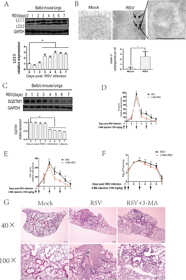
FIG 1.
RSV-induced autophagy enhances viral replication in the lungs of BALB/c mice. (A) BALB/c mice were mock infected or infected with RSV as described in Materials and Methods. Mouse lungs were harvested at indicated days and the expression of LC3II and GAPDH was analyzed with immunoblotting with specific antibodies. The relative levels of targeted proteins were quantitated by densitometry and normalized to GAPDH control. The data represent means ± SD for at least 3 mice per group. (B) BALB/c mice were treated as described for panel A for 5 days, and mice lungs were examined by transmission electron microcopy. The black arrows depict double-membraned autophagosomes. The bar graph represents the number of autophagosomes per cell. The data are from 50 cells per sample. Bar, 500 nm. (C) BALB/c mice were treated as described for panel A, and the expression of SQSTM1 and GAPDH of mice lungs was analyzed with immunoblotting with specific antibodies. The relative levels of targeted proteins were quantitated by densitometry and normalized to GAPDH control. The data represent means ± SD for at least 3 mice per group. (D and E) BALB/c mice were infected with RSV in the presence or absence of 3-MA as described in Materials and Methods. Mouse lungs were harvested at indicated days, and viral N (D) or NS1 (E) genes were detected by qRT-PCR. Gene expression was calculated by comparison to that of one mouse infected with RSV at day 1 postinfection (100%). Results are means ± SD for 5 mice per group. (F) BALB/c mice were treated as described for panels D and E, and lung homogenates at indicated days were prepared to detect viral titers according to the methods described in Materials and Methods. Results are shown as log10 PFU/g lung and represent means ± SD for 5 mice per group. (G) BALB/c mice were treated as described for panels D and E for 5 days, and lung pathology was detected by hematoxylin and eosin (H&E) stain. *, P < 0.05.

