FIG 2.
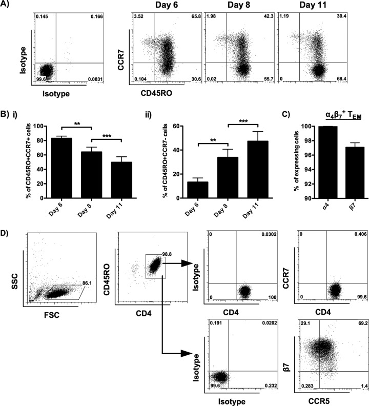
Characteristics of α4β7+ TEM isolated from α4β7+ MEMT. Purified CD4 T cells were cultured in the presence of gamma-irradiated RPMI8866 cells plus IL-2, IL-15, and RA. The phenotypes of the cells were analyzed by flow cytometry on day 6, day 8, and day 11 after CD45RO+ cell enrichment. (A) Representative flow cytometric analysis (donors = 8) shows the expression of CD45RO and CCR7 on allogeneic activated CD4 T cells. (B) Bar charts illustrate the kinetic changes in the percentage of CD45RO+ CCR7+ TCM (i) and CD45RO+ CCR7− TEM (ii) in allogeneic activated CD4 T cells (donors = 8). The phenotypes of α4β7+ TEM isolated from α4β7+ MEMT on day 11 were analyzed by flow cytometry. (C) Percentage of α4+ and β7+ α4β7+ TEM on day 11 (donors = 10). (D) Representative flow cytometric analysis (donors = 10) shows the expression of CD45RO, CCR7, integrin β7, and CCR5 on α4β7+ TEM. The positive cells were defined using the corresponding isotype controls. SSC, side scatter; FSC, forward scatter. Data are expressed as the mean ± SEM. **, P < 0.01; ***, P < 0.001.

