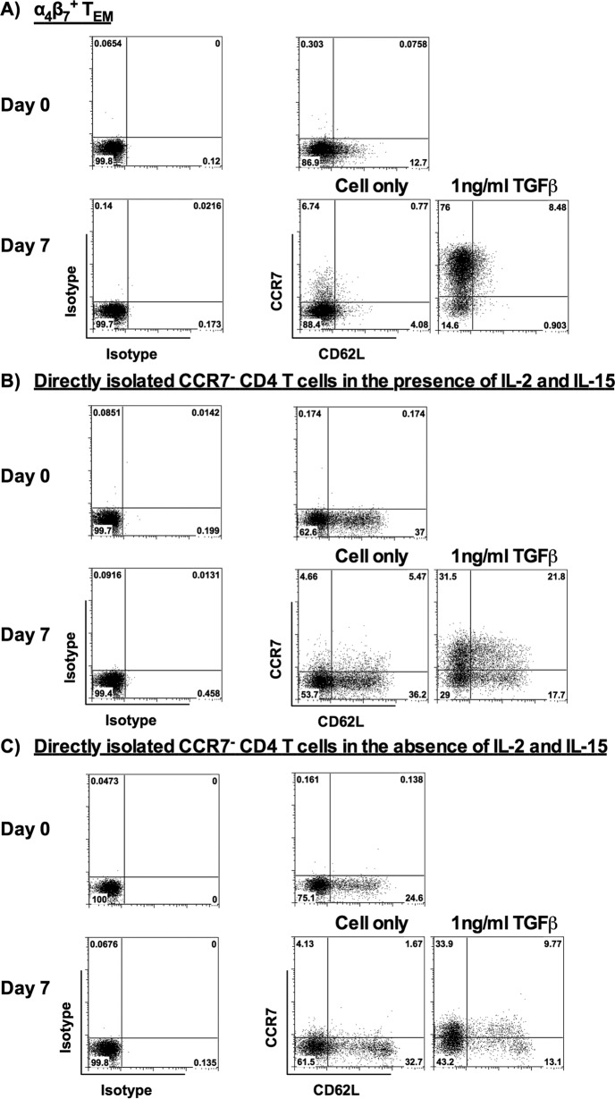
FIG 7.
Changes of CD62L expression on α4β7+ TEM and TEM directly isolated from peripheral blood in the presence or absence of TGF-β. (A) Representative flow cytometric analysis shows the expression of CCR7 and CD62L on the isolated α4β7+ TEM (day 0) and the α4β7+ TEM stimulated with 1 ng/ml TGF-β for 7 days (day 7) (donors = 8). (B and C) Representative flow cytometric analysis shows the expression of CCR7 and CD62L on CD3+ CD4+ CD45RA− CCR7− TEM directly isolated from peripheral blood on day 0 and on day 7 after stimulation with 1 ng/ml TGF-β in the presence (donors = 6) (B) or absence (donors = 6) (C) of IL-2 (10 ng/ml) and IL-15 (20 ng/ml). The positive cells were defined using the corresponding isotype controls.

