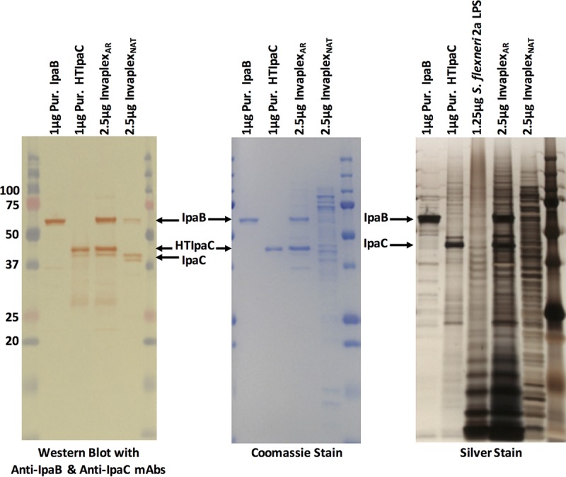
FIG 1 .

SDS-PAGE analysis of InvaplexAR and InvaplexNAT. Samples loaded were S. flexneri 2a InvaplexAR (2.5 µg), InvaplexNAT (2.5 µg), IpaB (1 µg), HTIpaC (1 µg), and purified S. flexneri 2a LPS (1.25 µg). Samples were separated by SDS-PAGE and blotted to nitrocellulose (left panel) or stained with Coomassie blue (middle panel) or silver (right panel). Nitrocellulose blots were probed with MAbs specific for IpaB (2F1) and IpaC (2G2). Silver-stained gels detected both proteins and LPS. Molecular mass standards are in the extreme left (Western blot and Coomassie blue) and right (Western blot and Coomassie blue and silver stain lanes) of the gels. Numbers at far left are kilodaltons.
