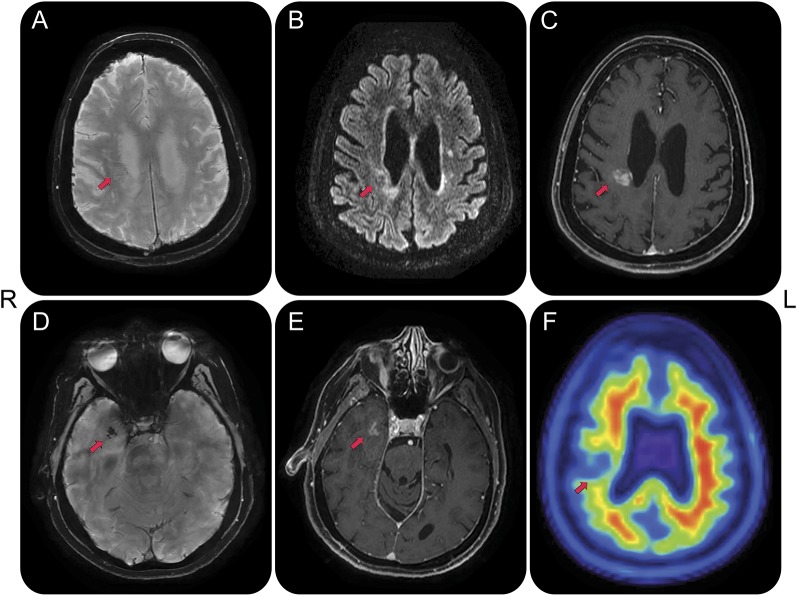
Figure 1. MRI and molecular amyloid PET.
MRI acquisitions obtained at age 68 years reveal 2 focal contrast-enhancing lesions in the right frontoparietal and temporal white matter and amyloid PET acquisition obtained at age 70 reveals decreased tracer uptake in the right frontoparietal lesion. Arrows point to lesions. (A) Susceptibility-weighted angiography sequence indicative of faint linear susceptibility artifact of the frontoparietal lesion, likely reflective of prominent vascularity, without features of microhemorrhages. (B) Fluid-attenuated inversion recovery sequence reveals hyperintense signal around the center of the lesion, possibly reflective of a combination of edema and white matter damage. (C) T1 with contrast sequence indicates blood–brain barrier disruption at the lesion center. (D) Susceptibility-weighted angiography sequence reveals confluent punctate susceptibility artifacts within the right temporal lesion. There were no distributed susceptibility artifacts other than the focal changes noted in the right anterior temporal lesion at age 68, which were absent at age 67. (E) Contrast enhancement of the right temporal lesion on T1 with contrast. (F) Amyloid [18F]florbetapir PET image reveals lack of cortical tracer uptake, making Alzheimer disease unlikely, and decreased tracer uptake at the lesion center, supportive of white matter breakdown, decreased perfusion, or lack of tracer sensitivity to specific amyloid pathology. There was no abnormal tracer uptake in the temporal lobe lesion. The temporal lesion remained stable, whereas the frontoparietal increased from 7 to 19 mm between ages 59 and 63, stabilizing thereafter. There was mild atrophy of the dorsolateral frontoparietal and medial temporal lobes, right worse than left (slices not shown).

