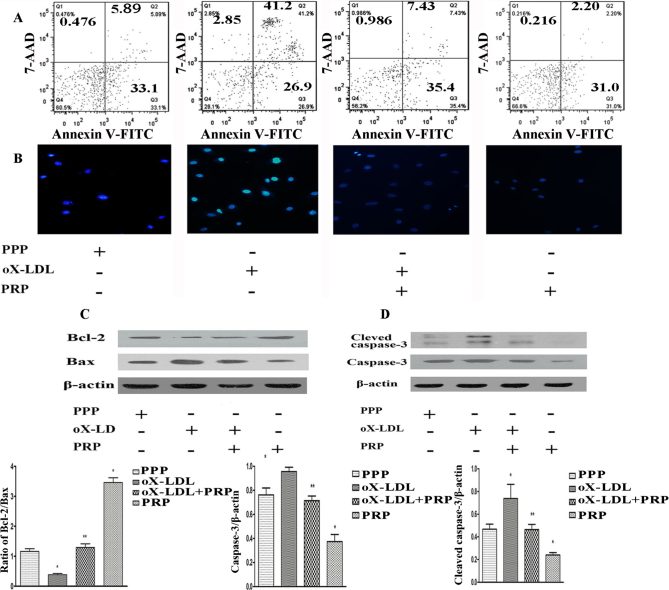
Figure 3.
Apoptotic cells and changes in death-related proteins were detected. A: Apoptotic cells were detected by flow cytometry. B: Cells emitting blue fluorescence were stained with Hoechst 33342. C: Bcl-2 and Bax were detected by western blotting. D: Cleaved caspase-3 and caspase-3 were detected by western blotting. *p<0.05 versus control; **p<0.05 versus oX-LDL.

