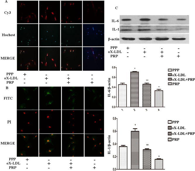
Figure 5.
IL-6 and IL-1 expression in four different groups. Secondary antibodies conjugated with Cy3 or FITC were used to detect IL-6 and IL-1, respectively. Cell nuclei were stained by Hochest 33342 and PI. A: IL-6 expression via immunofluorescence. B: IL-1 expression via immunofluorescence. C: IL-6 and IL-1 expression detected by western blotting (mean ± SD). *p<0.05 versus control; **p<0.05 versus oX-LDL.

