
Figure 3.
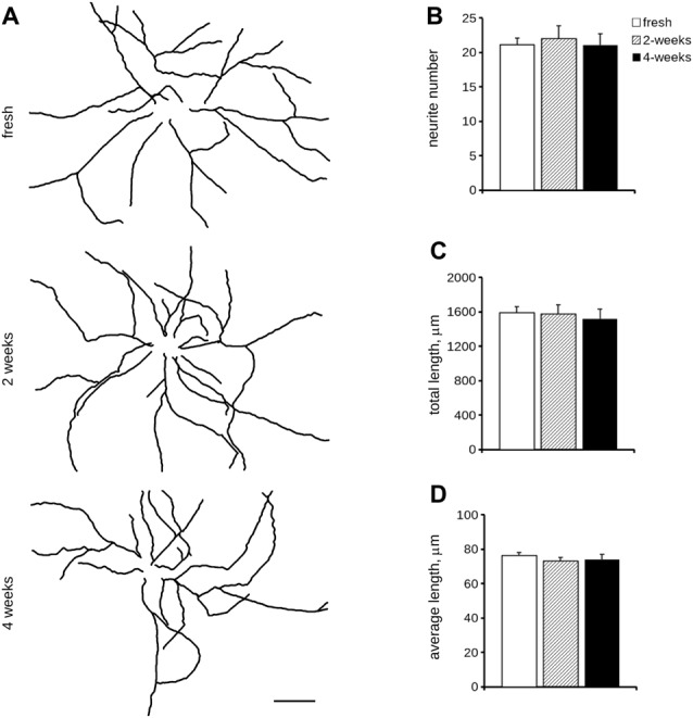
Morphological analysis of cryopreserved cultures. We analyzed morphology of acutely dissociated cortical neurons vs. cultures cryopreserved for 2 or 4 weeks. Neurons were infected at DIV4 and processed for immunofluorescence at DIV14. Panels show camera lucida tracing (A). Graphs show neurite number (B), total length (C) and average length (D). Data are reported as mean ± SEM, n = 15–22. Scale bar = 50 μm.