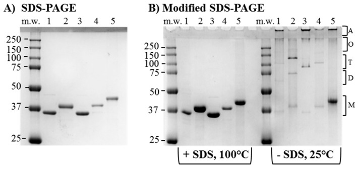
Figure 3.
Electrophoretic analysis of purified PorB/VDs. Coomassie staining of (A) Conventional SDS-PAGE. Lane 1: NL PorB (~34 kDa); Lane 2: PorB/VD1 (~37 kDa); Lane 3: PorB/VD2 (~35 kDa); Lane 4: PorB/VD3 (~37 kDa); Lane 5: PorB/VD4 (~38 kDa); (B) Modified SDS-PAGE. Left panel: samples dissolved in loading buffer with SDS and incubated at 100 °C for 5 min; Right panel: samples dissolved in SDS-free loading buffer and kept at 25 °C prior to electrophoresis Lane numbers are marked as in (A). The predicted positions of bands of molecular weight corresponding to monomers (M), dimers (D), trimers (T), oligomeric forms (O) and aggregates (A) are indicated by the brackets.

