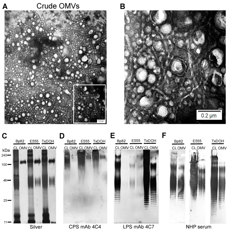
Figure 1.
Analysis of outer membrane vesicles (OMVs) and immunogenic polysaccharides. (A) Crude OMV negative stained TEM image showing numerous vesicles and flagellar material. (B) Zoom image of crude OMV from the area indicated by the white box in (A). (C) Silver stain of polysaccharide preparations from cell lysates (CL) and OMVs. (D) Western blot of the major B. mallei capsular polysaccharide (CPS) reveals the high molecular weight polysaccharide is present in all three biosafe strains and their OMVs. (E) Western blot of the LPS O-antigen from all three biosafe strains and their OMVs using B. mallei O-antigen specific mAb 4C7. (F) Reactivity of non-human primate (NHP) serum with cell lysate and OMVs from all three biosafe strains.

