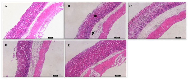
Figure 2.
Histological evaluation of the ethanol-induced gastric damage in mice. (A) Control stomach: intact gastric epithelium with organized glandular structure and normal submucosa could be seen; (B–E) ethanol-induced damage; (B) mice pre-treated with vehicle: * indicates damaged mucosal epithelium with disrupted glandular structure and arrow depicts oedema of submucosa and inflammatory infiltrate of mucosa; (C) Cm-SP 2 mg/kg; (D) Cm-SP 20 mg/kg; (E) Cm-SP 200 mg/kg. (C–E) depict a recovery in mucosa epithelium and reorganized glandular structure, as well as improvement of oedema by Cm-SP. (H&E staining; magnification 100×).

