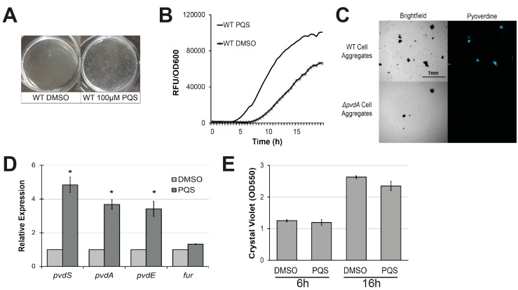
Figure 1.
Exogenous Pseudomonas quinolone signal (PQS) induces cell aggregation and promotes pyoverdine production. (A) Cell aggregate formation in P. aeruginosa PA14 treated with either dimethyl sulfoxide (DMSO) (left) or 100 µM PQS (right) after a 4 h growth period. (B) Pyoverdine fluorescence normalized to bacterial growth, measured over 24 h in P. aeruginosa treated with DMSO or 100 µM PQS. (C) Brightfield (left) or fluorescence (right) micrographs of pyoverdine expression in either wild-type PA14 (top) or PA14ΔpvdA, a pyoverdine biosynthesis mutant (bottom). Cell aggregates were visualized with a pyoverdine-specific fluorescence filter. (D) Expression of pyoverdine biosynthesis genes in bacteria treated with 100 µM PQS or DMSO after 6 h growth, as measured by quantitative, real-time PCR (qRT-PCR). Gene expression in PQS-treated bacteria was normalized to the solvent control. (E) Quantification of crystal violet staining of biofilm matrix from wild-type bacteria treated with either DMSO solvent or 100 µM PQS after 6 or 16 h growth. Crystal violet was solubilized in 30% acetic acid solution and quantified by absorbance at 550 nm. Error bars in (D) represent standard error of the mean (SEM) between three biological replicates. * Corresponds to p < 0.01 based on Student’s t-test. Pyoverdine production curves without normalization to bacterial growth are available in Figure S1.

