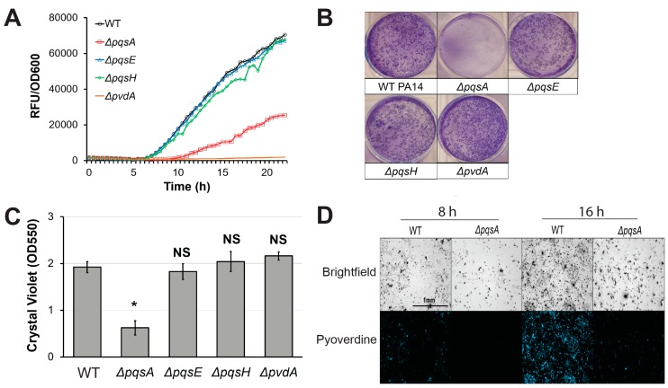
Figure 3.
PqsA is necessary for biofilm formation and pyoverdine production. (A) Pyoverdine fluorescence, normalized to bacterial growth, measured kinetically over 24 h in WT PA14 and PQS biosynthetic mutants. (B) Biofilm matrix of PQS biosynthetic mutants in 6-well plate stained with 0.1% crystal violet. (C) Quantification of crystal violet stain measured by absorbance at 550 nm after solubilizing in 30% acetic acid solution. (D) Brightfield (top) and fluorescence (bottom) micrographs of WT PA14 and ΔpqsA biofilm matrix cell aggregates visualized with a pyoverdine-specific fluorescence filter. Error bars in (B) represent SEM between three biological replicates. NS corresponds to p > 0.05, # corresponds to p < 0.05, and * corresponds to p < 0.01 based on Student’s t-test. Pyoverdine production curves without normalization to bacterial growth are available in Figure S1.

