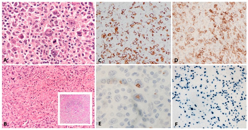
Figure 2.
EBV positive diffuse large B-cell lymphoma, not otherwise specified. (A) Polymorphous infiltrate of HRS-like cells in a background of lymphocytes and histiocytes (H&E, 400×). (B) Wide areas of necrosis are common and invasion of vascular walls is seen (inset) (H&E, 100×). (C) The majority of tumour cells are positive for CD20, which highlights markedly variable size of the lesional cells. (D) Most of the tumour cells are positive for CD30. (E) Occasional cells co-expressing CD15 are seen. (immunohistochemistry, C, D, 200×, E, 400×) (F) There is widespread positivity for EBER, which highlights variability in the size of the nuclei. (in-situ hybridization, 200×).

